History & Management of Short QT Syndrome

Diagnosis and Management of Short QT Syndrome patients

Abstract

Establishing a definition of Short QT Syndrome including symptomatology and QT interval duration is still a work in progress. It is clear, however, that SQTS is a rare, life-threatening, inherited heart disease presenting as SCD or aborted SCD in 34% and a family history of SCD in 15%. Genetic testing is important in diagnosing the disease, but so far with a causative mutation found in less than 25%. A benign variety of the disease has been observed in children with atrial fibrillation and a _KCNH2-V141M_ mutation, and just recently a mutation in the cardiac Cl,HCO3- exchanger AE3 was found to cause SQTS. Issues related to measuring and correcting the QT interval for heart rate has made it difficult to rely entirely on QT duration for the diagnosis of SQTS. In order to establish the diagnosis on firmer grounds, symptoms, family history and genetic testing need to be considered. While the benefit of implantation of an ICD as secondary prophylaxis against SCD in a patient with SQTS is well-documented, the benefit as primary prophylaxis is controversial and not proven by solid data. In two recent similar studies involving 115 patients at an approximate 5 year follow-up, an ICD implanted in 40 patients saved the lives of 12 who had presented with cardiac arrest in 11 and syncope in one. No appropriate shocks were delivered in any patients who did not have a history of either syncope or cardiac arrest. Currently Quinidine is the only drug which has undergone any clinical testing.

Dr. Preben Bjerregaard MD, DMSc

Dr. Preben Bjerregaard

MD, DMSc

Introduction

Short QT Syndrome (SQTS) is a very rare disease known for less than twenty years. The arrhythmic risk from a long QT interval had been discovered already in 19571 while the risk of a short QT interval was first noticed in 20002 when a sixteen-year-old female of Italian decent presented with new onset atrial fibrillation and QT/QTc intervals of 280/300 ms (Figure 1) at the same time as sudden cardiac death had been observed in a thirty-seven-year-old female from Spain with similar short QT- interval. In 2003 the family history of patients with short QT and familial sudden death was described by Gaita F, et al.3 Among the first 100 patients published with SQTS, 62 were from Western Europe (shortqtsyndrome.org). Early on following the discovery of SQTS the disease was poorly defined. In medical textbooks the normal for QT interval was usually given only by an upper limit and several large population studies were undertaken in order to define the lower limit of normal for the QT interval. But it was soon realized that just having a short QT interval by definition would not be enough to

Twelve-lead surface ECG (paper speed 25 mm/sec and 10 mm/mV) from 16-year-old girl who was the first person to be diagnosed with SQTS. She was later found to have a KCNH2 mutation. The QT interval is 280 msec at a heart rate of 68 beats/min. PQ-segment depression < 0.05 mV from the isoelectric TP-segment is seen in leads I, aVL and V2-V6.

Twelve-lead surface ECG (paper speed 25 mm/sec and 10 mm/mV) from 16-year-old girl who was the first person to be diagnosed with SQTS. She was later found to have a KCNH2 mutation. The QT interval is 280 msec at a heart rate of 68 beats/min. PQ-segment depression < 0.05 mV from the isoelectric TP-segment is seen in leads I, aVL and V2-V6.

qualify as SQTS and formulas like Bazett’s for correction of the QT interval for heart rate turned out to be especially inaccurate in patients with SQTS. It took however only four years after the first patient was diagnosed with SQTS until the first genetic mutation was found.4 Early on an implantable defibrillator was considered the best and possibly only option for prevention of SCD in patients with SQTS. QT interval prolonging drugs like Quinidine showed promising results. This review is based upon all SQTS papers published in English language medical journals 2000-2017 and will summarize experiences from diagnosing and managing SQTS patients, which is based more on clinical observations then statistically founded data. This review includes 220 patients (145 male) from 144 families with (SQTS).2-44 Many were published as case reports, but recent attempts especially in Europe (The European SQTS registry and The Inherited Arrhythmia Database in Pavia, Italy) to collect and present patient data from several centers together have made it possible to get a more complete picture of the disease. Recently a study of a large family in Denmark led to the discovery of a novel mechanism of SQTS in terms of a genetically reduced function of an anion exchanger.45

Dr. Preben Bjerregaard MD, DMSc

Dr. Preben Bjerregaard

MD, DMSc

Genetics in Short QT Syndrome

Out of the 144 families with Short QT Syndrome a total of 98 families (68%) have been genetically tested, and in 38 (39%), a causative gene-mutation has been identified. Since at least 1/3 of these were case reports of newly discovered mutations the chance of finding a causative mutation in a family with SQTS is in the neighborhood of 20 %. A total of 22 different mutations have been found in 9 different genes with 15 in single families.

Table 1:

Original study Gene Locus Nucleotie change Amino Acid Change Number of Published families Special Issues
Brugada R et al4 KCNH2 c1764a/c1764g N588K 6  
Sun Y. et al22 7q36.1 c1853t T618I 2  
Redpath CJ. et al16     E50D 1  
Harrell A.M. et al39   c1679>c I560T 1  
Akdis D. et al   c1891T>G S631A 1 new
Bellocq C. et al5 KCNQ1 g919c V307L 1  
Hong K.et al10 11p15.5   V141M 9  
Rhodes T.E. et al51     I274V 1 SIDS
Moreno C. et al40   t127910a F279I 1  
Mazzanti A. et al38     R259H 1  
Rothenberg I. et al44   c859G>A A287T 1  
Priori S.G. et al8 KCNJ2 g514a D172N 2  
Hattori T. et al26 17q24.3   M301K 1  
Deo M. et al.31   a896t E299V 2  
Ambrosini E. et al36     K346T 1 Twins
Mazzanti A. et al38 CACNA1C   R1977Q 1  
  12p13.3        
Templin C. et al24 CACNA2D1 c2264g S755T 1  
  7q21.11        
Thorsen K. et al45 SLC4A3 c1109G>A R370H 2  
  2q35        

In table 1 is shown the genetic findings in mutation positive patients with Short QT Syndrome.

Table 2:

Original study GENE locus Nucleotide change Amino acid change Number of published families
Antzelevitch C. et al13 CACNA1C c116t A39V 1
Antzelevitch C. et al13 12p13.3 a1468g G490R 1
Antzelevitch C. et al13 CACNA1C c1442t S481L 2
  10p12.33
Itoh H.et al18 KCNH2   R1135H 1
  7q36.1
Hong K et al28 SCN5A g2066a R689H 1
  3p21

Table 2 shows the genetic findings in mutation positive patients with a rare combination of overlapping ST-segment elevation, short QT interval and sudden cardiac death, first described by Antzelevitch C. et al in 2007.13 A clear difference between the two groups is in the penetrance of the mutations, with an almost 100% penetrance in SQTS patients. Incomplete penetrance in this group was only seen in the family with a KCNH2-E50D mutation.16 The penetrance in families in table 2 has been more like the 56% seen in patients with Brugada syndrome.47

Dr. Preben Bjerregaard MD, DMSc

Dr. Preben Bjerregaard

MD, DMSc

KCNQ1-V141M mutation positive patients with Short QT Syndrome

Among patients with potassium channel mutations, one group clearly separates from the others: patients with a KCNQ1-V141M mutation. This is the most frequent mutation in patients with SQTS, found in 10 (8 female) patients from 9 families sharing a similar clinical picture, starting as fetal bradycardia in 7, bradycardia at birth in 2 and as slow atrial fibrillation at the age of three years old in 1.10,32,33,39,42,43 The QT and QTc intervals were all very short, varying between 200 and 290 msec. All patients subsequently developed slow atrial fibrillation which was either resistant to DC cardioversion or offered only a temporary correction after atrial fibrillation was replaced by sinus bradycardia or junctional rhythm, suggesting sinus node dysfunction. All 10 patients ended up in persistent atrial fibrillation and due to a slow heart rate of 40-50 beats per minute or symptomatic pauses, a pacemaker was implanted in 5, including 1 who developed cardiomyopathy and the only one who died during follow-up. Patients without a pacemaker did well despite a heart rate of 40-60 beats per minute. In 3 patients where genetic testing of the parents was performed, results were negative and there was no family history of similar disease except in one case where a patient’s father had a KCNQ1-V141M mutation and a history of chronic atrial fibrillation since the age of 3 years old .39 His ECG available in supplemental material shows atrial fibrillation with a short QTc.

Dr. Preben Bjerregaard MD, DMSc

Dr. Preben Bjerregaard

MD, DMSc

Genetically reduced function of an anion exchanger as a novel mechanism

Based upon the fact that cation channel mutations explain < 25 % of SQTS cases and other classes of genes therefore likely   to be associated with the development of this disease, Thorsen K et al.45 very recently in a study of two Danish families with dominantly inherited SQTS specifically looked for involvement of other genes by whole exome sequencing. In their study they identified a novel genetic etiology for SQTS in terms of a mutation in the anion exchanger Solute Carrier Family 4 Member 3 (SLC4A3) gene, which encodes a Cl,HCO3 –exchanger (AE3). The AE3 transports Cl- into the cardiomyocyte in exchange for transport of HCO3- out of the cell. The mutation leads to a trafficking defect, decreased Cl,HCO3-exchange over the cell membrane, increased pHi in combination with a decrease in Cl-I and was shown to shorten QT duration in zebrafish embryos.

A unique feature of the anion channel mutation study is the size of family 1 consisting of 42 screened members with 23 carriers of the SLC4A3 c.1109 G>A variant. The index patient had presented at age 31 years old with cardiac arrest during sleep and two relatives had died suddenly and unexplained at the age of 41 and 42 years, respectively. All carriers had a QT ≤ 360 ms, and a QTc ≤ 370 ms (mean 340 +/- 18 ms) and 19 non-carriers all   had a QTc > 370 msec (mean 402 +/- 24 msec) (p< 0.001). In an unrelated family consisting of 6 members included for verification there were 4 carriers of the SCL4A3 c1109 mutation who all had QTc < 370 msec. The proband in this family had died suddenly and unexpectedly at rest 22 years old.

The authors have clearly identified a novel mutation in the cardiac chloride-bicarbonate exchanger AE3 in two independent families with SQTS, and in zebrafish demonstrated the impact of the mutation on duration of systolic contraction and on QTc duration. The authors suggest that the mechanism through which a defect in Cl,HCO3-exchange leads to shortening QTc likely relates to a combined effect of a decrease in HCO3- efflux (i.e. increased pHi) and a decrease in Cl- influx, both affecting the repolarization. It is also suggested that these findings in addition to offering insight into mechanisms of arrhythmia in general may provide new treatment possibilities in some patients with SQTS. (Data from this study are not included anywhere else in this paper except in Table 1).

Regarding the studies of cation channel gene mutations one concern has been that large families (> 6 affected with SQTS) have never been described and thus genetic findings have been based on biological plausibility rather than strong genetic segregation or linkage data.

Dr. Preben Bjerregaard MD, DMSc

Dr. Preben Bjerregaard

MD, DMSc

Clinical presentation of SQTS families

The famous medical maxim, “You see what you look for and you recognize what you know,” may express one reason why so few patients until now have been diagnosed with SQTS. To find patients with SQTS you must know when to suspect it, and this is where the clinical presentation of published data can be helpful. In 110 out of the 140 families in the literature was the clinical presentation revealed:

Table 3. Clinical presentation of 110 probands out of 144 SQTS families in the literature where this information was available. 

Clinical presentation Number of probands % af families
SCD or aborted SCD 37 34
Family history of SCD 17 15
Syncope 17 15
Atrial fibrillation and/or bradycardia in newborn 11 10
Atrial fibrillation in a child 4 4
Ventricular tachycardia 2 2
Autism-Epilepsy 1 1
Paroxysmal atrial fibrillation in an adult 1 1
Routine ECG 20 18

Unfortunately, in half of the families the diagnosis of SCD in a close relative comes too late to benefit the patient in relation to the threat of their own event.  Syncope has occurred in a fair number of cases, but due to the common occurrence of this symptom, its relationship to SQTS can be difficult to establish. A newborn with atrial fibrillation and a slow heart rate, or atrial fibrillation in a child should always be met with high suspicion of SQTS. The high number of SQTS families found by routine ECG (17 out of 47 (36%)) in the study based upon the inherited Arrhythmia Database in Pavia, Italy38 compared to only three in all other studies is puzzling, but may have something to do with the higher prevalence of SQTS in Italy than in other countries. Another explanation may be Italy’s strong program of pre-participation screening, where all young people 12 years of age or older who engage in competitive sport activities are required to undergo ECG and exercise stress testing, providing significantly more data on Italians.

Dr. Preben Bjerregaard MD, DMSc

Dr. Preben Bjerregaard

MD, DMSc

Suggested recommendations on SQTS diagnosis

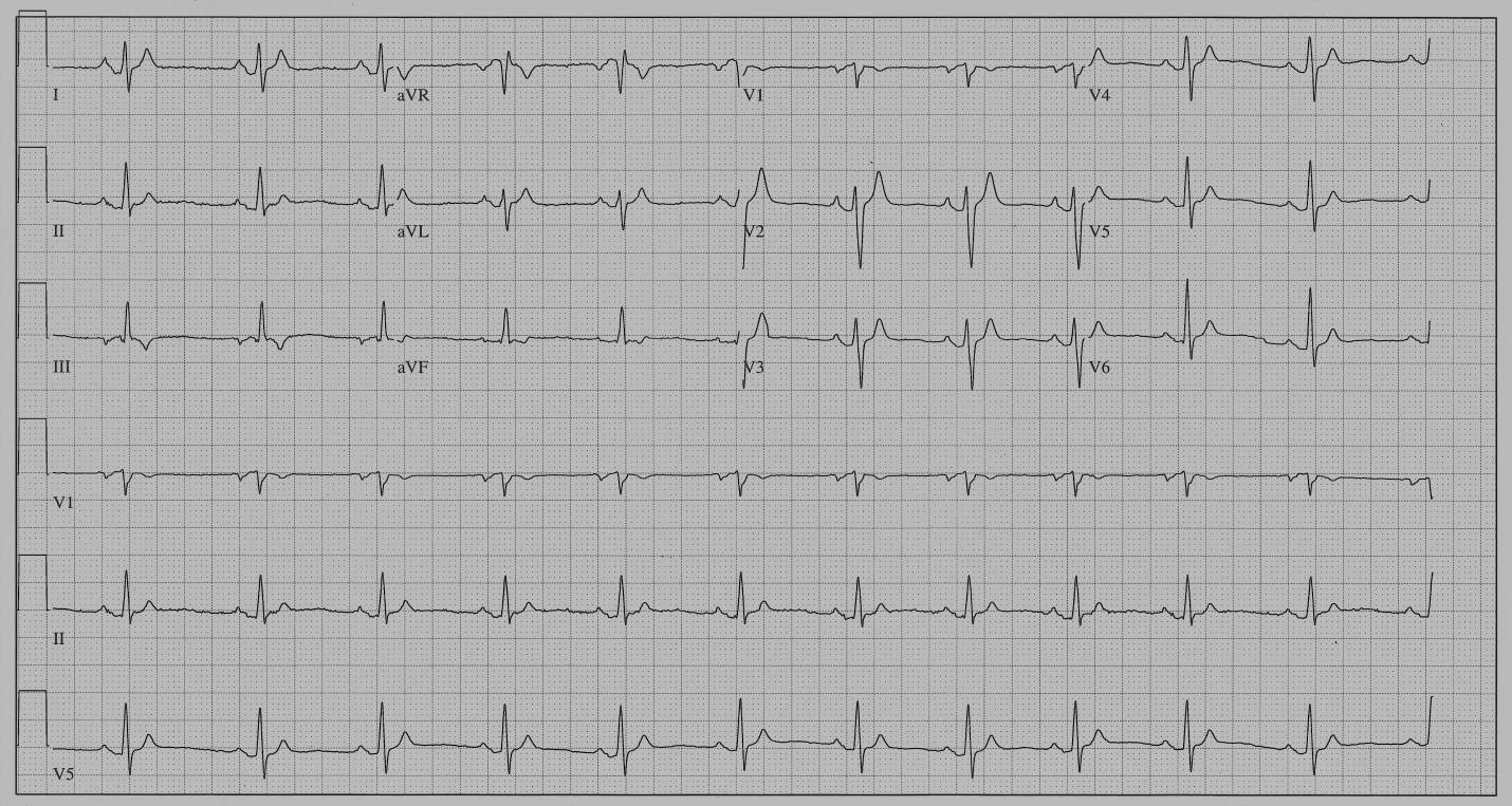
Regarding the second part of the maxim, “you recognize what you know,” there are several issues regarding the QT-interval that are important to be aware of when making the diagnosis of SQTS. As shown by exercise testing of patients with SQTS, the QT interval alters very little with changes in heart rate. A recent exercise test study from five European Centers of 21 patients with SQTS (QT: 276 +/- 27 ms) and 20  healthy subjects (QT: 364 +/- 25 ms) found a QT-HR slope in SQTS patients of -0.53+/- 0.15 ms compared to -1.29 +/- 0.30 ms in healthy subjects.46  Because of a difference between the two groups, not only in terms of SQTS, but also short QT intervals, it is difficult to tell whether the variations in HR-QT slopes are due to the difference in QT interval duration, or SQTS. It may therefore be too soon to fully accept the authors suggestion, that, “in subjects with QTc intervals between 340 and 360 ms, the presence of a QT/HR relationship slope under -0.9 ms.beat/min could help distinguishing affected subjects from healthy individuals.”   Until a comparison of patients with SQTS and healthy individuals with similar QT-intervals have been studied we cannot be certain whether the HR-QT slope can be used as an aid in diagnosing SQTS.  An important consequence of the diminished effect of heart rate on the QT-interval in patients with a short QT interval is that correction calculations based on Bazett’s formula will overcorrect short QT intervals to a much higher degree than in people with normal QT intervals. At slow HR’s this may lead to over-diagnosing SQTS, and at fast HR’s to under-diagnosing SQTS.

 Despite these well-documented shortcomings in using QTc  especially in patients with SQTS, QTc is commonly used in recommendations on SQTS diagnosis. In order to avoid extreme overcorrections by this method it is, however, recommendable to obtain an ECG at a heart rate as close to 60 bpm as possible (repeat ECG, Holter) and limit the use of Bazett’s correction formula in diagnosing SQTS to heart rates between 50 and 70 bpm. It is also unrealistic to expect that a single QTc value will distinguish all cases of SQTS from healthy individuals. Since the HRS/EHRA/APHRS Expert Consensus Statement on the Diagnosis and Management of Patients with SQTS from 2013,47 there has been two major studies with new information about SQTS suggesting that an update in criteria for diagnosing SQTS would be appropriate.  Prior to 2013 only 5 of 70 SQTS patients with information of individual QTc’s had a QTc > 330 ms .  Since then two multicenter trials including 126 patients,23,38 have used  QTc ≤ 340 ms as stand-alone criterion for making the diagnosis of SQTS. In one of these studies where individual QTc’s were given,38 21 out of 73 patients, of whom 84% were male, had QTc > 330 ms  while all of them had a QTc ≤ 360 ms. Based upon these data it might be appropriate to use QTc ≤ 340 ms as stand-alone criterion for SQTS. In addition, based upon recent data from the largest SQTS family published so far,    where four (all males) out of twenty-three mutation-positive patients (17%) had a QTc ≥ 360 ms, the upper limit for  QTc should probably be 370 ms  instead of 360 ms when SQTS is diagnosed in the presence of one or more of the following: a pathogenic mutation, family history of SQTS, family history of SCD at age 40, survival of a VT/VF episode in the absence of heart disease, atrial fibrillation at a young age and syncopal episode that strongly suggests a cardiac arrhythmia. In comparison to the 2013 guidelines atrial fibrillation at a young age has been added based upon the discovery of KCNQ1-V141 mutation positive children with atrial fibrillation, and the addition of syncopal episodes is based upon three multicenter studies21,23,38 which includes the majority of patient data published to date. In the first study syncope was not defined, in the second study syncope had to be of arrhythmic origin and in the third study syncope was defined as a transient loss of consciousness in the absence of alternative explanations (e.g., labyrinthitis, orthostatic hypotension, or clear, vagally mediated events).

Dr. Preben Bjerregaard MD, DMSc

Dr. Preben Bjerregaard

MD, DMSc

Special features in the ECG in patients with SQTS

In addition to the short QT interval there are certain other features in the ECG in patients with SQTS which appear to occur with a higher prevalence than expected by chance alone. One such finding is PQ Segment Depression (PQD), defined as ≥ 0.05 mV PQ depression from the isoelectric TP segment. Beginning with the first patient discovered with SQTS (Figure 1),  PQD was noticed, and when a study of 64 patients with SQTS in the European SQTS registry was carried out in 2015, 48 PQD was found in 52 (81%), compared to only 24% in a control group of 117 age- and sex-matched subjects. The best ECG lead in which to identify PQD in was lead II, followed by V3 and aVF.

Another common feature in patients with SQTS is tall and peaked T waves independent of genotype. In some SQTS patients a biphasic negative-positive T wave immediately following the QRS has been observed, replacing a normal ST-segment, while in other SQTS patients the ST-segment is missing altogether.49

 Finally, early repolarization has been observed in the ECG of 65% of the SQTS patients compared to only 30% of a control group with short QT interval, and only 10% of a normal QT control cohort.21

EP studies in patients with SQTS have shown short refractory periods both in atria and ventricles, as well as demonstrating a peculiar tendency to induce VF when placing an electrode catheter in the heart, but in general, EP studies are not very helpful in establishing the diagnosis of SQTS or predicting cardiac arrest

A proposed scoring evaluation for SQTS put forward by Gollob et al,50 in analogy with the Schwartz score for the LQTS, has been difficult to validate due to the low number of patients with SQTS, but been useful in bringing clinicians together on the agreement of what constitutes the diagnosis of SQTS. A modified score system has in addition shown some potential in predicting arrhythmic events in children with SQTS.30

Dr. Preben Bjerregaard MD, DMSc

Dr. Preben Bjerregaard

MD, DMSc

Management of Short QT syndrome

Based upon the family history of those individuals with SQTS where several members died suddenly, often at a young age, early studies of patients with SQTS suggested a very high risk of SCD. An implantable cardioverter-defibrillator (ICD) was considered the first choice therapy, but alternative therapy was necessary, especially in small children and in adults who did not want an ICD. The first clinical results from testing of Quinidine as a QT-prolonging and sudden-death preventative drug in patients with SQTS appeared as early as 2006.11 Over the years, the drugs Ibutilide, Flecainide, Sotalol, Disopyramide, Nifekalant, Propafenone, Carvedilol, Metoprolol and Amiodarone have been considered as alternatives to Quinidine, which has, at times, been difficult to obtain and often has intolerable side-effects, but none have undergone serious clinical testing, primarily due to the scarcity of patients and the low event rate in SQTS patients suitable for clinical trials.  Quinidine is currently the only antiarrhythmic drug which has undergone some degree of clinical testing in such patients and only considered potentially beneficial in patients where the drug was able to prolong and preferably normalize the QT interval. In a recent multicenter trial of 53 patients from the European SQTS registry,23 hydroquinidine (HQ) was tested in 22 subjects. In 6 cases, it had to be discontinued because of poor therapeutic compliance; in 2 cases because of no effect on the QT interval; and in two additional cases where 2 subjects reported gastrointestinal side effects. Twelve patients (8 adults and 4 children) received HQ for a mean period of 76 +/- 30 months. During an electrophysiologic study at baseline, 7 patients had induced VF, whereas none had VF induced after HQ. During a follow-up of approximately five years, none of the 12 patients on HQ had any arrhythmic events. However, the event rate for the entire group of 53 patients was very low with only 2 episodes of VF. 

An ICD is still considered the best option as secondary prophylaxis in patients with SQTS following cardiac arrest, whereas its role in primary prophylaxis is less clear. The results from two very similar multicenter studies23,38  with a combined 115 patients at an approximate 5 year follow-up, are very important in this debate. An ICD implanted in 40 patients saved the lives of 12 who had episodes of VT/VF. Eleven of those 12 patients had presented with cardiac arrest. The remaining patient who survived had initially presented with syncope. In addition, one was saved by an external defibrillator and only one patient, who had refused an ICD, died.  No appropriate shock was delivered in any patient who did not have a history of either syncope or cardiac arrest.

Dr. Preben Bjerregaard MD, DMSc

Dr. Preben Bjerregaard

MD, DMSc

Risk stratification of patients with Short QT Syndrome

Due to the low number of patients with SQTS, and given the fact the longest follow-up study to date has been approximately 5 years, it is not possible to present any concrete assessment of the risk individuals born with SQTS face of sudden death, but Mazzanti et al38 estimated the cumulative probability of experiencing a first occurrence of cardiac arrest by 40 years of age as 41%, with the highest risk occurring during the first year of life and between ages 20 and 40 years. Patients with a KCNQ1-V141M mutation, however, are an exception to the high risk of death during the first year of life. Of 11 known patients with this mutation, the only one who had cardiac arrest in childhood was a patient with SQTS and cardiomyopathy, possibly occurring as a complication to pacemaker implantation.

Electrophysiological testing has not been useful in predicting cardiac arrest. A clear relationship between clinical risk and QT/QTc duration in patients with SQTS has not been proven, but a strong trend to increased events based on shorter absolute QT or QTc intervals was demonstrated in the original scorecard paper by Gollob MH et al50 and in the paper by Villafane J et al30 in children. In the paper with long-term follow up of patients with SQTS by Giustetto C et al23 the QTc values in a population with short QT did not, however, distinguish between asymptomatic subjects and those with cardiac arrest.23

 The only predictor of cardiac arrest in a patient with SQTS found so far has been a previous history of cardiac arrest. The risk of cardiac arrest in healthy subjects was assessed in a Finnish population study of 10,822 subjects age 44 +/- 8.4 years.52 During follow-up for 29 +/- 10 years, 43 subjects, or 0.4% of the population who had a QTc < 340 msec (and therefore by current definition SQTS), none experienced sudden cardiac death, aborted sudden cardiac death, or documented ventricular tachyarrhythmias, which illustrates how rare SQTS is, and how low the risk of cardiac arrest is in people with SQTS, based upon an incidental finding of a short QT. Any benefit from implantation of an ICD in such individuals is very questionable. More long-term follow-up of asymptomatic people with a family history and/or genetic predisposition is needed to assess their risk and possible benefit of an ICD.

Dr. Preben Bjerregaard MD, DMSc

Dr. Preben Bjerregaard

MD, DMSc

Conclusion

Due to the persistent commitment at a few centers with special interest in studying SQTS both in the laboratory and clinically, our knowledge about the disease less than 20 years after it was discovered is substantial, but several key questions still needs to be answered. We still need to find ways to prevent more people from dying suddenly due to undiagnosed SQTS, and improve our ability to risk stratify people with a short QT interval. Limited testing of Quinidine in patients with SQTS has shown promising results, but due to the difficulties in using and getting the drug, clinical testing of other antiarrhythmic drugs needs to be pursued. In light of the newly discovered anion gene mutation there is a need for reevaluation of the pool of SQTS patients with hitherto negative genetic test results and to include anion gene mutations in future genetic testing for SQTS.

Dr. Preben Bjerregaard MD, DMSc

Dr. Preben Bjerregaard

MD, DMSc

References

  1. Jervell AL, Lange-Nielsen F. Congenital deaf-mutism, functional heart disease with prolongation of the QT interval and sudden death. Am Heart J 1957;54:59-68
  2. Gussak I, Brugada P, Brugada J, Wright S, Kopecky SL, Chaitman BR, Bjerregaard, P. Idiopathis Short QT Interval: A New Clinical Syndrome? Cardiology 2000;94:99-102
  3. Gaita F, Giustetto C, Bianchi F, Wolpert C, Schimpf R, Riccardi R, Grossi S, Richiardi E, Borggrefe M. Short QT Syndrome. A Familial Cause of Sudden death. Circulation 2003;108:965-970
  4. Brugada R, Hong K, Dumaine R et al. Sudden Death Associated With Short-QT Syndrome Linked to Mutations in HERG. Circulation 2004;109:30-35
  5. Bellocq C, van Ginneken CB, Bezzina CR, Alders M, Escande D, Mannens MMAM, Baró I, Wilde AAM. Mutation in the KCNQ1 Gene Leading to the Short QT-Interval Syndrome. Circulation 2004;109:2394-2397
  6. Riera ARP, Ferreira C, Dubner SJ, Schapachnik E, Soares JD, Francis J. Brief Review of the Recently Described Short QT Syndrome and Other Cardiac Channelopathies. A.N.E. 2005;(10)3;371-377
  7. Maury P, Hollington L, Duparc A, Brugada R. Short QT Syndrome: Should we push the frontier forward? Heart Rhythm 2005;2:1135-1137
  8. Priori SG, Pandit SV, Rivolta I et al. A Novel Form of Short QT Syndrome (SQT3) Is Caused by a Mutation in the KCNJ2 Circ Res. 2005;96:800-607
  9. Kirilmaz A, Ulusoy RE, Kardesoglu E. Ozmen N, Demiralp E. Short QT Interval: a case report. Journal of Electrocardiology 2005;38:371-374
  10. Hong K, Piper DR, Diaz-Valdecantos A et al. De Novo KCNQ1 mutation responsible for atrial fibrillation and short QT syndrome in utero. Cardiovasc Res. 2005;68:433-440
  11. Giustetto C, Di Monte F, Wolpert C, Borggrefe M, Schimpf R, Sbragia P, Leone G, Maury P, Anttonen O, Haïssaguerre M, Gaita F. Short QT Syndrome: clinical findings and diagnostic-therapeutic implications. Eur Heart J 2006;27:2440-2447
  12. Lu LX, Zhou W, Zhang X, Cao Q, Yu Kanglong, Zhu Changqing. Short QT Syndrome: A case report and review of literature. Resuscitation 2006;71:115-121
  13. Antzelevitch C, Pollevick GD, Cordeiro J et al. Loss-of-Function Mutations in the Cardiac Calcium Channel Underlie a New Clinical Entity Characterized by ST-Segment Elevation, Short QT Intervals, and Sudden Cardiac Death. Circulation 2007;115:442-449
  14. Mizobuchi M, Enjoji Y, Yamamoto R, Ono T, Funatsu A, Kambayashi D, Kobayashi T, Nakamura S. Nifekalant and Disopyramide in a patient with Short QT Syndrome: Evaluation of Pharmacological Effects and Electrophysiological Properties. PACE 2008;31:1229-1232
  15. Fichet J, Genee O, Pierre B, Babuty D. Fatal QT interval. Am J Emerg Med 2008;26:739e5-e6
  16. Redpath CJ, Green MS, Birnie DH, Gollop MH. Rapid genetic testing facilitating the diagnosis of short QT syndrome. Can J Cardiol 2009;25:e133-5
  17. Villafane J, Young ML, Maury P, Wolpart C, Anttonen O, Hamilton R, Kannankeril PJ, Fischbach PS. Short QT Syndrome in a Pediatric Patient. Pediatr Cardiol 2009;30:846-850
  18. Itoh H, Sakaguchi T, Ashihara T et al.  A novel KCNH2 mutation as a modifier for short QT interval. Int. J. Cardiol 2009;137:83-85
  19. Efremides M, Letsas KP, Weber R, Gavrielatos G, Filippatos GS, Sideris A, Kardaras F. Recurrent syncope associated with a distinct ECG pattern consisting of short QT interval, early repolarization and atrioventricular block. Clin Res Cardiol 2009;98:807-810
  20. Sun Y, Zhang P, Li X, Guo J. Inappropriate ICD Discharge Due to

T-Wave Oversensing in a Patient with Short QT Syndrome. PACE 2010;33:113-116

  1. Watanabe H, Makiyama T, Koyama T et al. High prevalence of early repolarization in short QT syndrome. Heart Rhythm 2010;7:647-652
  2. Sun Y, Quan X-Q, Fromme S, Cox RH, Zhang P, Zhang L, Guo D, Guo J, Patel C, Kowey PR, Yan G-X. A novel mutation in the KCNH2 gene associated with short QT syndrome. JMCC 2011;50:433-441
  3. Giustetto C, Schimpf R, Mazzanti A, Scrocco C, Maury P, Anttonen O, Probst V, Blanc J-J, Sbragia P, Dalmasso P, Borggrefe M, Gaita F. Long-Term Follow-up of Patients With Short QT Syndrome. J Am Coll Cardiol 2011;58:587-95
  4. Templin C, Ghadri J-R, Rougier J-S et al. Identification of a novel loss-of-function calcium channel mutation in short QT syndrome (SQTS6). Eur Heart J 2011;32(9):1077-1088
  5. Chinushi M, Sato A, Izumi D, Furushima H. Nifekalant Enlarged the Transmural Activation-Recovery Interval Difference as Well as the Peak-to-End Interval on Surface ECG in a patient with Short-QT Syndrome. J Cardiovasc Electrophysiol 2012;23:877-880
  6. Hattori T, Makiyama T, Akao M, Ehara E, Ohno S,.Iguchi M, Nishis Y, Sasaki K, Itoh H, Yokode M, Kita T, Horie M, Kimura T. A novel gain-of-function KCNJ2 mutation associated with short-QT syndrome impairs inward rectification of Kir2.1 currents. Cardiovascular Research 2012;93(4):666-673
  7. Bun S-S, Maury P, Giustetto C, Deharo J-C. Electrical Storm in Short-QT Syndrome Successfully Treated with Isoproterenol. J Cardiovasc Electrophysiol 2012;23(9):1028-30
  8. Hong K, Hu J, YU J, Brugada R. Concomitant Brugada-like and short QT electrocardiogram linked to SCN5A European Journal of Human Genetics 2012;20:1189-1192
  9. Babaoğlu K, Binnetoğlu K, Altun G, Tuzcu V. A 13-year-old boy with a short QT interval. Anatolian Journal of Cardiology 2012;12(3);274-275
  10. Villafane J, Atallah J, Gollob MH et al. Long-Term Follow-up of a Pediatric Cohort With Short QT Syndrome. J Am Coll Cardiol 2013;61:1183-91
  11. Deo M, Ruan Y, Pandit SV, Shah K, Berenfeld O, Blaufox A, Cerrone M, Noujaim SF, Denegri M, Jalife J, Priori SG. KCNJ2 mutation in short QT syndrome 3 results in atrial fibrillation and ventricular proarrhythmia. PNAS 2013;110(11):4291-96
  12. Maltret A, Wiever-Vacher S, Denis C, Extramiana F, Morisseau-Durand MP, Fressart V, Bobbet D, Chabbert C. Type 2 short QT syndrome and ventricular dysfunction: Mirror of the Jervell and Lange-Nielsen syndrome. Int J Cardiol 2014;171(2):291-3
  13. Villafane J, Fischbach, Gebauer R. Short QT Syndrome Manifesting with Neonatal Atrial Fibrillation and Bradycardia. Cardiology 2014;128:236-240
  14. Sadeghian S, Bozorgi A, Safkhani Z. Short QT syndrome and idiopathic ventricular tachycardia in a 28-year-old young man: a potential disease-specific link? Europace 2014;16(11):1645
  15. Portugal G, Oliveira MM, Cunha PS, Ferreira F, Lousinha A, Fiarresga A, Nogueira da Silva M, Ferreira RC. Short QT syndrome presenting as syncope: How short is too short? Rev Port Cardiol 2014;33(10):649.e1-.e6
  16. Ambrosini E, Sicca F, Brignone MS et al. Genetically induced dysfunctions of Kir2.1 channels: Implications for short QT3 syndrome and autism-epilepsy phenotype. Human Molecular Genetics 2014;23(18)4875-4886
  17. Suzuki H, Hoshina S, Ozawa J, Sato A, Minamino T, Aizawa Y, Saitoh A. Short QT Syndrome in a boy diagnosed on screening for heart disease. Pediatr Int 2014;56(5):774-76
  18. Mazzanti A, Kanthan A, Monteforte N et al. Novel Insight Into the Natural History of Short QT Syndrome. J Am Coll Cardiol 2014;63:1300-8
  19. Harrell DT, Ashihara T, Ishikawa T, Tominaga I, Mazzanti A, Takahashi K, Oginosawa Y, Abe H, Maemura K et.al. Genotype-dependent differences in age of manifestation and arrhythmia complications in short QT syndrome. International Journal of Cardiology 2015;190:393-402
  20. Moreno C, Oliveras A, de la Cruz A. A new KCNQ1 mutation at the S5 segment that impairs its association with KCNE1 is responsible for short QT syndrome. Cardiovascular research 2015;107:613-623
  21. Ergül Y, Özyilmaz I, Onan SH, Güzeltaş Short QT syndrome on a 14 year-old patient: The first pediatric case from Turkey. Anatol  J Cardiol 2015;15:586-91
  22. Righi D, Silvetti MS, Drago F. Sinus bradycardia, junctional rhythm, and low-rate atrial fibrillation in Short QT syndrome during 20 years of follow-up: three faces of the same genetic problem. Cardiology in the Young 2016;26:589-592
  23. Sarguella-Brugada G, Campuzano O, Iglesias A, Grueso J, Bradley DJ, Kerst G, Shmorhun D, Brugada J, Brugada R. Short QT and atrial fibrillation: A KCNQ1 mutation-specific disease. Late follow-up in three unrelated children. Heart Rhythm Case Reports 2015;1(4):193-7
  24. Rothenberg I, Piccini I, Wrobel E, Stallmeyer B, Müller J, Greber B, Strutz-Seebohm N, Schulze-Bahr E, Schmitt N, Seebohm G. Structural interplay of Kv1 and KCNE1 is essential for normal repolarization and is compromised in short QT syndrome 2 (Kv7.1-A287T). Heart Rhythm Case Reports 2016;2:521-529
  25. Thorsen K, Dam VS, Kjær-Sørensen K et al. Loss-of-activity-mutation in the cardiac chloride-bicarbonate exchanger AE3 causes short QT syndrome. Nat Commun 2017;8(1):1696. doi: 10.1038/s41467-017-01630-0.
  26. Giustetto C, Scrocco C, Schimpf R et al. Usefulness of exercise test in the diagnosis of short QT syndrome. Europace 2015;17:628-634
  27. Priori SG, Wilde AA, Horie M et al. HRS/EHRS/APHRS Expert Consensus Statement on the Diagnosis and Management of Patients with Inherited Primary Arrhythmia Syndromes. Heart Rhythm 2013;10(12);1933-1963
  28. Tülümen E, Guistetto C, Wolpert C et al. PQ Depression in Short QT Syndrome Patients: A Novel Marker for Diagnosing Short QT Syndrome. Heart Rhythm 2014;11(6);1024-1030
  29. Pérez-Riera AR, Paixã-Almeida A, Barbosa-Barros R, Yanowitz FG, Baranchuk A, Dubner S, Chagas ACP. Congenital short QT syndrome: Landmarks of the newest arrhythmogenic cardiac channelopathy. Cardiol J 2013;20(5):464-471
  30. Gollop MH, Redpath CJ, Roberts JD. The Short QT Syndrome. Proposed Diagnostic Criteria. J Am Coll Cardiol 2011;57:802-812
  31. Rhoades TE, Abraham RA, Welch RC et al. Cardiac Potasseum Channel Dysfunction in Sudden Infant Death Syndrome. J Moll Cell Cardiol 2008;44(3):571-581

Anttonen O, Junttila MJ, Rissanen H, Reunanen A, Viitasalo M, Huikuri HV. Prevalence and Prognostic Significance of Short QT Interval in a Middle-Aged Finnish Population. Circulation 2007;116:714-720

Dr. Preben Bjerregaard MD, DMSc

Dr. Preben Bjerregaard

MD, DMSc